Articles
- Page Path
- HOME > Korean J Community Nutr > Volume 28(3); 2023 > Article
- Review
- Mercury exposure is associated with obesity: a systematic review and meta-analysis
- Jimin Jeon, Kyong Park
-
Korean Journal of Community Nutrition 2023;28(3):192-205.
DOI: https://doi.org/10.5720/kjcn.2023.28.3.192
Published online: June 30, 2023

2Professor, Department of Food and Nutrition, Yeungnam University, Gyeongsan, Korea

-
Corresponding author:
Kyong Park, Tel: +82-53-810-2879, Fax: +82-53-810-4666,
Email: kypark@ynu.ac.kr
- 177 Views
- 19 Download
- 1 Crossref
- 0 Scopus
Abstract
Objectives
Previous studies have evaluated the association between mercury exposure and obesity but have yielded mixed conclusions. The aim of this study was to systematically review and summarize scientific evidence regarding the association between mercury exposure and obesity in the human population.
Methods
We conducted a systematic search of PubMed, Web of Science, Scopus, and Science Direct for articles related to mercury exposure and obesity. Meta-analyses of the highest and lowest categories of mercury levels were evaluated using a random effects model. Begg’s test was used to detect publication bias.
Results
A total of 9 articles were included. The pooled random effects odds ratio (OR) for mercury exposure and obesity of all 9 studies was 1.66 (95% confidence interval [CI]: 1.16-2.38). This positive association was evident in adults (OR: 1.61, 95% CI: 1.02-2.54) and among studies with Asian populations (OR: 2.00, 95% CI: 1.53-2.59), but not among those with North America/African populations (OR: 0.90, 95% CI: 0.50-1.65).
Conclusions
The present meta-analysis identified a positive association between mercury exposure and obesity. These findings suggest that toxic environmental metals such as mercury may be an important risk factor for obesity along with dietary habits and lifestyles.
Published online Jun 30, 2023.
https://doi.org/10.5720/kjcn.2023.28.3.192
Mercury exposure is associated with obesity: a systematic review and meta-analysis
Abstract
Objectives
Previous studies have evaluated the association between mercury exposure and obesity but have yielded mixed conclusions. The aim of this study was to systematically review and summarize scientific evidence regarding the association between mercury exposure and obesity in the human population.
Methods
We conducted a systematic search of PubMed, Web of Science, Scopus, and Science Direct for articles related to mercury exposure and obesity. Meta-analyses of the highest and lowest categories of mercury levels were evaluated using a random effects model. Begg’s test was used to detect publication bias.
Results
A total of 9 articles were included. The pooled random effects odds ratio (OR) for mercury exposure and obesity of all 9 studies was 1.66 (95% confidence interval [CI]: 1.16-2.38). This positive association was evident in adults (OR: 1.61, 95% CI: 1.02-2.54) and among studies with Asian populations (OR: 2.00, 95% CI: 1.53-2.59), but not among those with North America/African populations (OR: 0.90, 95% CI: 0.50-1.65).
Conclusions
The present meta-analysis identified a positive association between mercury exposure and obesity. These findings suggest that toxic environmental metals such as mercury may be an important risk factor for obesity along with dietary habits and lifestyles.
Introduction
Mercury is a highly toxic heavy metal [1] that affects all living creatures, including plants and animals, by natural or artificial processes [2]. Inorganic mercury flows into water, where it is converted into methyl mercury by microorganisms. It then accumulates in small aquatic creatures, which are eventually consumed by larger fishes such as sharks, whales, and tuna [3]. Ingested mercury bioaccumulates readily in the human body, where it affects the central nervous system and kidney function [4, 5] and can cause chronic diseases such as cardiovascular disease and hypertension [6].
The results of several studies have suggested that exposure to mercury may increase the risk of developing a range of cardiovascular and metabolic diseases. For example, the Coronary Artery Risk Development in Young Adults study found that higher levels of toenail mercury were linked to an increased risk of diabetes [7]. Similarly, research conducted in Europe and Israel demonstrated a positive correlation between toenail mercury levels and myocardial infarction [8]. Furthermore, a Finnish study revealed that the accumulation of mercury in hair and urine was associated with an increased risk of acute myocardial infarction, coronary artery disease, and cardiovascular disease mortality [9]. A recent meta-analysis also showed that hair mercury levels were positively associated with hypertension, with a U-shaped relationship between mercury levels and hypertension risk [10]. Collectively, these findings suggest that exposure to mercury may pose significant health risks, highlighting the importance of monitoring and regulating mercury exposure in various settings.
Recent research suggests that exposure to mercury may be associated with obesity or abdominal obesity [11, 12, 13, 14, 15, 16, 17, 18, 19], which is a well-known risk factor for various chronic diseases and mortality [20]. Most epidemiological studies on this topic were conducted in Korea, possibly due to the availability of blood mercury concentration data from national surveys or the cultural interest in fish and seafood. The mechanism by which mercury exposure affects obesity is thought to involve interference with endocrine signals and carbohydrate and lipid metabolism, leading to obesity development [21, 22, 23]. However, some studies, such as the National Health and Nutrition Examination Survey (NHANES) and the Modeling of Epidemiologic Transition Study, have not found significant links between blood mercury levels and obesity, or have even shown the opposite trend, where higher mercury exposure is associated with lower levels of obesity [24, 25, 26, 27]. In light of the contradictory findings, it is important to conduct a meta-analysis to thoroughly investigate the potential association between mercury exposure and obesity. Such an analysis can help refine our understanding of the potential health risks of mercury exposure and shed light on the reasons for the conflicting results seen in previous studies.
The main objective of this study was to conduct a systematic review and meta-analysis of the latest research literature on the association between mercury exposure and obesity. Furthermore, the research also aimed to investigate the heterogeneity between different studies and provide insights into potential sources of variation across the results. By achieving these objectives, this study aimed to contribute to a better understanding of the association between mercury exposure and obesity, which could have significant implications for public health policy and clinical practice.
Methods
1. Search Strategy
A systematic review and meta-analysis of studies on the association between mercury and obesity were performed in accordance with the Preferred Reporting Items for Systematic Reviews and Meta-Analyses guideline [28]. PubMed, Web of Science, Scopus, and Science Direct databases were searched for articles related to mercury and obesity published from January 1990 to October 2022. The keywords used were (“mercury” or “methyl mercury”) and (“body mass index” or “BMI” or “obesity” or “obese” or “overweight” or “abdominal obesity” or “weight” or “waist circumference” or “waist-hip-ratio” or “waist hip ratio” or “WHR” or “adiposity” or “body fat”). Based on the population, intervention, comparison, outcomes, and study design (PICOS) criteria, the inclusion criteria were defined as shown in Table 1.
Table 1
PICOS criteria for inclusion and exclusion of studies
2. Study selection
Duplicate articles were deleted using ENDNOTE 20 (Thomson Reuters, San Francisco, CA, USA). The titles and abstracts of the remaining articles were then reviewed according to the criteria for selection of articles. If study inclusion eligibility could not be determined through the title and abstract, the full text was reviewed before a decision on inclusion. Originally two investigators (J.J. and K.P.) independently screened studies according to the inclusion and exclusion criteria of study selection, and additional five investigators (J.I., C.K., U.J., H.Y., Y.P.) reviewed and updated the selected studies. The inclusion criteria were as follows: 1) original research; 2) observational study designs (including cross-sectional, case-control, nested-case control, and cohort designs); 3) mercury as the major exposure factor (as measured by blood, hair, urine, or toenail mercury concentrations); 4) obesity as the major outcome (as measured by BMI, WC, obesity, or abdominal obesity); and 5) published in either English or Korean. The exclusion criteria were as follows: 1) non-original articles such as letters, reviews, or comments; 2) not conducted in humans; and 3) insufficient information to calculate the effect size for the association between mercury and obesity or abdominal obesity (odds ratio [OR] / prevalence ratio [PR] or 95% confidence interval [CI]). The results of the literature search and selection process are presented in Fig. 1.
Fig. 1
Flow chart of study selection for the meta-analysis
3. Quality assessment
We performed a quality assessment of the included studies using the Quality Assessment Tool for Observational studies checklist [29]. The tool consisted of 14 questions concerning study objectives, participant eligibility, study power, definition of exposure and outcome measures, any potential sources of bias, timing between exposure and outcome, and other factors related to internal validity. Study quality was scored by summing the total number of applicable questions classified as “yes”, and then rated as good, fair, or poor by the investigators. All included studies were rated as good or fair (Supplementary Table 1).
4. Statistical Analysis
In this meta-analysis, the effect size between mercury levels and obesity from each study was aggregated using the OR/PR and 95% CI of the highest vs. lowest mercury levels with a random effect model, which considers the between-study heterogeneity. Except for Fig. 2, which stratifies the pooled odds ratios of the association between mercury levels and both obesity and abdominal obesity, we maintained a single-outcome approach for our meta-analysis. When a study reported both obesity and abdominal obesity, we included only obesity to avoid over-representation, given its higher frequency of reporting across studies. Heterogeneity between studies was assessed using the Higgin’s I2 statistic and Cochrane’s Q test. Heterogeneity was defined as an I2 > 50% or a P-value < 0.1 in the Cochrane’s Q test [30]. To identify the potential sources of heterogeneity, a subgroup analysis was conducted. Furthermore, publication bias was evaluated using Begg's and Mazumdar's rank correlation test and funnel plot test. An influence analysis was also performed using the leave-one-out method. This meta-analysis was performed using STATA version 13.1 (Stata Corp LP, College Station, TX, USA).
Results
1. Literature search
A total of 11,692 articles were identified from PubMed, Web of Science, Scopus, and Science Direct and 2 articles were additionally identified through manual searching of citation lists and related articles (n = 2). Of these, duplicates (n = 4,928) and articles that met the exclusion criteria or were unrelated to the topic (n = 6,719) were excluded. Among the 47 remaining articles, those with overlapping datasets and those without an OR/PR or 95% CI were excluded (n = 38). Finally, nine articles were included in the meta-analysis. The characteristics of the nine studies are shown in Table 2. Altogether, 40,044 participants from 8 different cross-sectional studies and 1 case-control study were included in the meta-analysis. Most studies (7 of 9) investigated adults, whereas two studies investigated children and adolescents. All studies evaluated the accumulation of mercury in blood or toenail samples, but most studies estimated the association between blood mercury levels and obesity without classifying sex. The individual studies were carried out in Korea (n = 5), China (n = 1), and countries from North America or Africa (n = 3), respectively. Two studies reported the prevalence of obesity, two studies reported the prevalence of abdominal obesity, and five studies reported both the prevalence of obesity and abdominal obesity.
Table 2
Characteristics of studies included in the meta-analysis
2. Pooled results on the association between mercury exposure and obesity
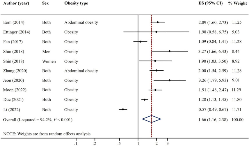
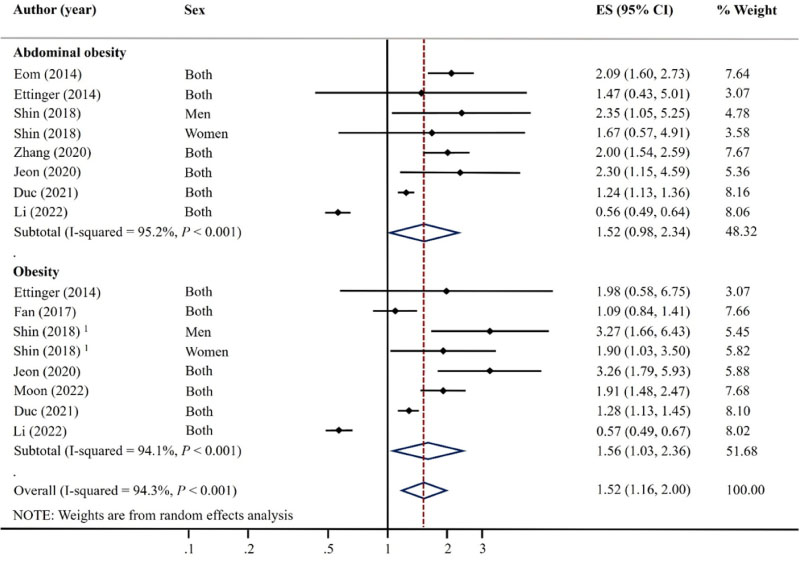
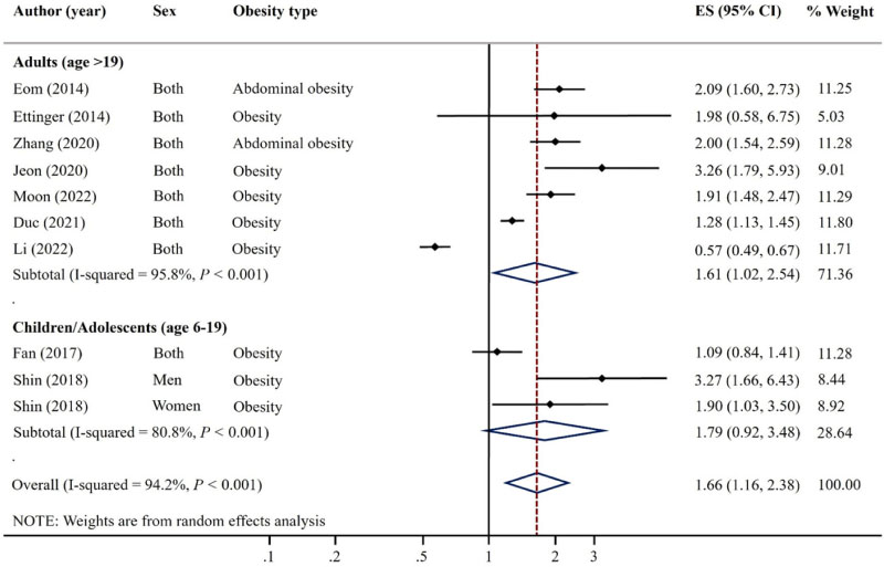
Fig. 2 shows the results of pooling studies on the association between mercury exposure and obesity. We observed a positive association between mercury exposure and obesity (OR: 1.66, 95% CI: 1.16-2.38), with a high between-study heterogeneity (I2 = 94.2%). Subgroup analyses of the association between mercury exposure and obesity according to obesity type showed that mercury exposure was positively associated with obesity, defined by BMI (I2 = 94.1%, OR: 1.56, 95% CI: 1.03-2.36), but not in the abdominal obesity (I2 = 95.2%, OR: 1.52, 95% CI: 0.98-2.34) (Fig. 3). However, in a subgroup analysis by continent in which the studies were conducted, heterogeneity was reduced in both Asia and North America or Africa, but a positive association between mercury exposure and obesity was only shown in Asia (I2 = 80.6%, OR: 2.00, 95% CI: 1.53-2.59), but not in North America or Africa (I2 = 90.3%, OR: 0.90, 95% CI: 0.50-1.65) (Fig. 4). In the analysis stratified by age, a positive association between mercury exposure and obesity was observed in adults (I2 = 95.8%, OR: 1.61, 95% CI: 1.02-2.54) but not in children or adolescents (I2 = 80.8%, OR: 1.79, 95% CI: 0.92-3.48) (Fig. 5).
Fig. 4
Forest plot of the pooled odds ratios of the association between mercury levels and obesity stratified by continent [14, 15, 16, 17, 18, 19, 24, 25, 26]. *Ettinger (2014) study included participants from countries in Africa (Ghana, South Africa, and Seychelles) as well as North America (Jamaica and the USA). ES, effect size; CI, confidence interval
3. Influence analysis and publication bias
We conducted a leave-one-out analysis to assess the influence of each study on the summary OR. We found that no single study significantly affected the pooled effect size (supplementary Fig. 1). Analysis of publication bias using funnel plots showed an asymmetric trend at the lower right of the funnel plot. However, Begg’s and Mazumdar’s test showed no publication bias (Begg’s test = 0.418).
Discussion
This study investigated the association between mercury exposure and obesity using a systematic review and meta-analysis. As a result, this study found a positive association between mercury exposure and obesity. However, a significant positive association was observed in Asia, but not in North America or Africa. In addition, the association was evident in adult population.
White adipose tissue (WAT) is involved in regulating energy storage and maintaining homeostasis [31], and produces various adipokines, such as adiponectin and leptin, which affect glucose, lipid metabolism, and immune response [32]. Dysfunction in WAT can cause dysregulation of adipokine secretion, leading to obesity. Several experimental studies have reported that mercury may act as a WAT disruptor [23, 33, 34]. An in vivo study investigating the effects of mercury on WAT revealed an abnormal decrease in adipocyte size due to the excess increase in the size of the smallest adipocytes. As a result, leptin mRNA levels in WAT and blood leptin levels were decreased [34]. Lack of leptin, an adipocyte hormone regulating obesity, can induce obesity [35]. In addition, mercury exposure can interfere with peroxisome proliferator-activated receptor-α (PPARα) and peroxisome proliferator-activated receptor-γ (PPARγ) mRNA expression levels in adipocytes [33]. The dysregulation of PPARα and PPARγ expression can cause the accumulation of triglycerides and disorders controlling glucose, eventually leading to obesity [36]. Long-term chronic mercury exposure to low doses may reduce adipocyte size, plasma insulin levels, glucose tolerance, and increase plasma glucose and triglyceride levels [23]. In addition, mercury may inhibit 3β-hydroxysteroid dehydrogenase production, which is involved in the synthesis and secretion of sex hormones. It can also reduce estrogen and testosterone formation, which affects body fat accumulation [37, 38, 39]. However, the mechanism underlying the effect of mercury on obesity in humans is poorly understood, and further research is needed to understand this relationship.
In our meta-analysis, we found that the association between mercury exposure and obesity varied between countries. Specifically, when analyzing data from adult participants in the NHANES, the study results showed an inverse association between blood mercury levels and obesity [25]. However, when analyzing data from children and adolescents in NHANES, the results showed no significant association [26]. In contrast, the majority of other studies, mostly conducted in Asian countries, demonstrated a positive association between mercury exposure and obesity. Li et al. [25] stated that the mechanism underlying the link between mercury exposure and obesity remains unclear, although an animal study showed that HgCl2 treatment can decrease serum leptin levels and down-regulate leptin mRNA expression in white adipose tissue, resulting in reduced adipocyte size [34]. However, cumulative studies from Asia, especially in Korea, have consistently reported a positive association between mercury exposure and obesity using various biomarkers (such as blood and toenail samples) and datasets (including national and regional data).
These divergent findings may be attributed to differences in the range and levels of mercury exposure in the body. The average blood mercury levels of adults were 0.5 µg/L and 0.85 µg/L in North America or Africa (respectively) [24], while in Korea it was 3.50 µg/L [40]. Children and adolescents in Korea also showed the highest blood mercury levels [15, 26]. Indeed, the average blood mercury levels of children and adults in Asia have been shown to be higher than those of other countries [41, 42]. Considering that the general association between nutritional intake and disease risk shows a U-shaped curve [43], the effects of mercury toxicity on health outcomes may be less observable in countries with lower levels of blood mercury exposure due to insufficient variability. The most common mercury exposure method in humans occurs through food, especially seafood. Asia’s high levels of mercury in the body are likely due to frequent intake of fish and shellfish [44]. In addition, larger fish tend to contain higher levels of mercury due to bioaccumulation [45]. Asia is the biggest fish supplier and consumer in the world. In particular, Korea is the biggest seafood consumer, with an annual intake of over 54.8 kg of seafood per person between 2013 and 2015, which is higher than in any other countries [46]. Unlike in the North America, where large predatory fish such as tuna, salmon, and cod are predominantly consumed [47], Koreans consume various kinds of fish, including relatively small-sized fish such as mackerel and anchovies as well as large predator species [48]. According to a study analyzing the correlation between consumption of various fish and blood mercury levels, higher intake of mackerel and anchovies was positively associated with the levels of blood mercury, suggesting that small-sized fish as well as large predator fish may affect the accumulation of mercury in the body [48]. Therefore, Asia, especially Koreans with their higher levels of mercury accumulation in the body, may be influenced by a higher intake of seafood, regardless of seafood size.
Despite the potential risks associated with mercury exposure, it is important to note that fish is generally considered a healthier alternative protein source to red meat due to its lower energy content and higher unsaturated fatty acids. This dietary preference might contribute to generally leaner physique observed in Asian populations, despite higher mercury levels, and presents a complex interplay between dietary habits, environmental exposure, and health outcomes. Understanding this balance between the nutritional benefits of fish consumption and potential mercury toxicity risks is crucial, especially for populations with high seafood intake. Further research is necessary to fully understand these relationships and develop dietary guidelines that can maximize health benefits while minimizing potential risks.
Selenium may also have contributed to the meta-analysis results. Selenium is an essential trace element with antioxidant function [49], which dilutes mercury toxicity [50]. A study in Estonia showed that hair mercury levels decreased significantly after administration of selenium supplements [51]. It was also found that selenium modifies the association between mercury and chronic diseases [52, 53]. A study of European populations with low levels of selenium in the body showed that higher mercury levels were associated with cardiovascular disease [8, 54], whereas no such association was found in Americans with relatively high selenium levels [55]. A study conducted in Korea revealed a significant positive association between toenail mercury levels and metabolic syndrome and dyslipidemia at lower levels of toenail selenium. In contrast, this association was attenuated and became non-significant at higher levels of toenail selenium [52, 53]. Selenium levels in food vary greatly, as they are affected by the levels of selenium in the cultivated soil [56], which differ by region and country. Korean selenium levels in soil were reported as 0.03-0.24 mg/kg [57], which was lower than in the United Kingdom (0.1-4.0 mg/kg), Spain (0.17-0.39 mg/kg), or the United States (< 0.1-4.3 mg/kg), and the overall average level (0.4 mg/kg) [58, 59]. Blood selenium levels in each country also show identical trends with selenium soil levels in each country [60]. Thus, different levels of selenium in the body may modify the association between mercury and obesity.
The association between mercury exposure and obesity also differed by age. However, few studies have analyzed this association in children and adolescents, and even the countries where the investigations were conducted showed differences in blood mercury levels, so it is difficult to explain how age difference affected the association between mercury and obesity. Thus, further studies are needed to investigate the effect of mercury on obesity in diverse age groups.
This study had several limitations that should be noted. The meta-analysis incorporated studies that were adjusted for multiple potential confounding factors. However, the studies’ observational nature suggests that there might be residual confounding factors that were not measured or are unknown. Given that all included studies were cross-sectional and case-control designs, it is important to acknowledge that these study designs, by their nature, could potentially introduce a risk of reverse causality. Besides, the limited geographic regions covered by the relatively small number of studies may have contributed to considerable between-study heterogeneity resulting from differences between the study populations, potentially decreasing the strength of the evidence obtained from the pooled analyses. The meta-analysis included a subgroup analysis based on country to investigate potential sources of heterogeneity. However, the consideration of ethnicity or race would have been crucial in discussing dietary culture and genetic background, which could significantly influence the association between mercury exposure and obesity. Regrettably, these variables were not available for the meta-analysis as studies did not report results based on race or ethnicity, especially in studies with mixed races, such as those conducted in the United States. Furthermore, most studies did not have available information on the effect of selenium, which is an essential potential confounding variable or effect modifier in the association between mercury exposure and obesity. As a result, the potential heterogeneity caused by selenium could not be adequately addressed in the meta-analysis. Finally, there was insufficient data to perform a dose-response meta-analysis to investigate the effects of mercury on obesity accurately. It is crucial since the range of mercury exposure may vary by population. More studies with larger sample sizes and diverse populations are necessary to confirm the findings and explore potential sources of heterogeneity further.
Conclusion
In conclusion, this meta-analysis showed that a high level of mercury exposure was associated with higher prevalence of obesity in Asia, whereas this association was not evident in North America or Africa. Our findings suggest that exposure to heavy metals such as mercury may play an important role in the development of obesity, along with dietary and lifestyle factors. Given these findings, it may be worth considering public health strategies aimed at reducing exposure to environmental contaminants like mercury as part of a multi-faceted approach to preventing and managing obesity. However, due to the observational nature of the included studies, a causal relationship between mercury and obesity cannot be established with certainty. Therefore, further research in the form of prospective cohort studies and clinical trials, conducted across various countries, are necessary to provide higher-quality evidence on this association.
Supplementary Materials
Risk of bias assessment of included studiesSupplementary Table 1
Influence analysis of pooled odds ratios, depicting meta-analysis estimates with the omission of each named studySupplementary Fig. 1
Conflict of Interest:As the Editor-in-Chief of the Korean Journal of Community Nutrition, I, Kyong Park, declare a potential conflict of interest with this publication. I ensured neutrality by abstaining from the manuscript's review process, managed by an independent associate editor. Otherwise, there are no financial or other issues that might lead to a conflict of interest.
Funding:This research was supported by the National Research Foundation of Korea grant funded by the Korea government (grant number: NRF-2021R1A2C1007869). The funding sponsor had no role in the study design; collection, analysis, and interpretation of data; writing of the report; and the decision to submit the article for publication.
Acknowledgments
The authors thank Jihyun Im, Unhui Jo, Chaehyun Kim, Hyeonji Yoo, and Yeeun Park for their technical contributions to this work.
Data Availability
Data sharing is not applicable to this article as no new data were created or analyzed in this study.
References
-
Risher JF. In: World Health Organization. Elemental mercury and inorganic mercury compounds: Human health aspects. Geneva, Switzerland: World Health Organization; 2003. pp. 7-9.
-
-
Bernhoft RA. Mercury toxicity and treatment: A review of the literature. J Environ Public Health 2012;2012
-
-
Park YK, Seok KJ, Lee SH, Jang SC, Jung WS, Lee JM. Association between GFR and hair mercury, lead level. Korean J Fam Med 2015;5(3):961–965.
-
-
Duruibe JO, Ogwuegbu M, Egwurugwu J. Heavy metal pollution and human biotoxic effects. Int J Phys Sci 2007;2(5):112–118.
-
-
Hoffman DJ, Heinz GH. Effects of mercury and selenium on glutathione metabolism and oxidative stress in mallard ducks. Environ Toxicol Chem 1998;17(2):161–166.
-
-
National Heart Lung and Blood Institute. Quality assessment tool for observational cohort and cross-sectional studies [internet]. National Heart Lung and Blood Institute; 2013 [cited 2022 Sep 15].Available from: https://www.nhlbi.nih.gov/health-
topics/study- quality- assessment- tools .
-
-
Vachhrajani KD, Chowdhury AR. Distribution of mercury and evaluation of testicular steroidogenesis in mercuric chloride and methylmercury administered rats. Indian J Exp Biol 1990;28(8):746–751.
-
-
United Nations Environment Programme. Global Mercury Assessment 2018 [internet]. United Nations Environment Programme; 2018 [cited 2022 Feb 6].Available from: https://www.unenvironment.org/resources/publication/global-
mercury- assessment- 2018 .
-
-
Zillioux EJ. Mercury in fish: History, sources, pathways, effects, and indicator usage. In: Armon RH, Hanninen O, editors. Environmental Indicators. Berlin: Springer; 2015. pp. 743-766.
-
-
Food and Agriculture Organization of the United Nations. The State of World Fisheries and Aquaculture 2016 [internet]. Food and Agriculture Organization of the United Nations; 2016 [cited 2022 Oct 18].
-
-
European Market Observatory for Fisheries and Aquaculture Products. The EU fish market: 2018 edition [internet]. European Market Observatory for Fisheries and Aquaculture Products; 2018 [cited 2022 Nov 14].Available from: https://www.eumofa.eu/market-
analysis#yearly .
-
-
Virtanen JK, Voutilainen S, Rissanen TH, Mursu J, Tuomainen TP, Korhonen MJ, et al. Mercury, fish oils, and risk of acute coronary events and cardiovascular disease, coronary heart disease, and all-cause mortality in men in eastern Finland. Arterioscler Thromb Vasc Biol 2005;25(1):228–233.
-
-
Choi YS, Hesketh JE. Nutritional biochemistry of selenium. J Korean Soc Food Sci Nutr 2006;35(5):661–670.
-
-
Xing K, Zhou S, Wu X, Zhu Y, Kong J, Shao T, et al. Concentrations and characteristics of selenium in soil samples from Dashan Region, a selenium-enriched area in China. Soil Sci Plant Nutr 2015;61(6):889–897.
-
-
Holland HD, Lollar BS, Turekian KK. In: Environmental geochemistry. Oxford, UK: Elsevier; 2005. pp. 46-48.
-

 KSCN
KSCN


 Cite
Cite


